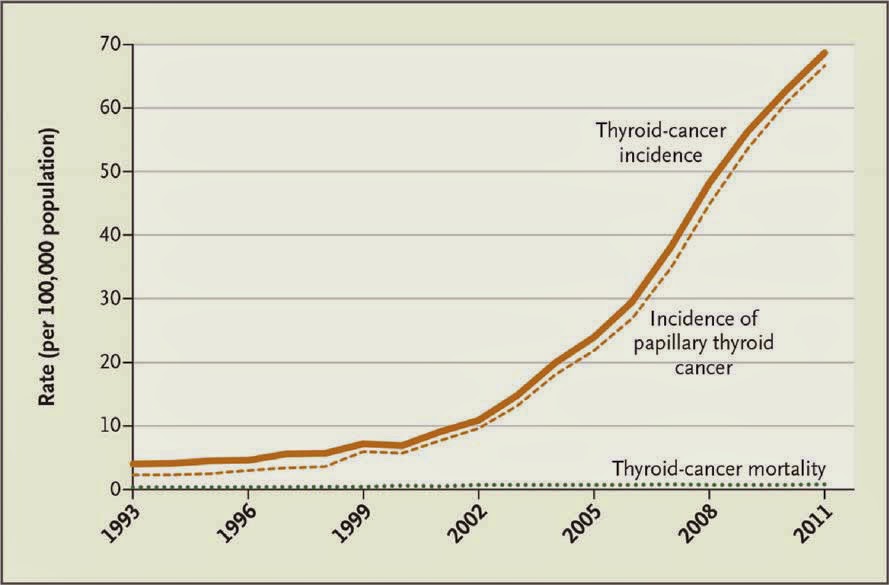
Trends
in Thyroid Cancer Incidence and Mortality in the United States, 1974-2013
Korea’s Thyroid-Cancer “Epidemic” — Screening and Overdiagnosis (and wireless phone use?)
November 5, 2014
According to today’s issue of the New England Journal of Medicine, South Korea has experienced a thyroid cancer epidemic in recent years (see paper and Figure below).
“Thyroid cancer is now the most common type of cancer diagnosed in South Korea.”
The authors of this paper attribute the “epidemic” to a government-sponsored cancer screening program. As evidence, they report,
That widespread screening identifies more cancer is not surprising. This could at least partly explain the increasing incidence of thyroid cancer observed in South Korea, and nine other countries including the U.S.
The authors argue that most of these cancers are not life-threatening and advise other countries against widespread screening for thyroid cancer:
Rather, I would like to focus on the question why has thyroid cancer become so prevalent in at least ten nations? According to the American Cancer Society, although some thyroid cancers are linked to exposure to ionizing radiation, “the exact cause of most thyroid cancers is not yet known.”
—
Is mobile phone use contributing to increased incidence of thyroid cancer?
July 9, 2014
The incidence of thyroid cancer has been increasing rapidly in recent years in many countries including the U.S., Canada, and Israel.
A headline in Haaretz a year ago March reads, “
Israeli scientists find possible link between cellphone use, thyroid cancer.”
In response to questions posed to me on this topic today from several individuals, I did a PubMed search. Although I did not find any epidemiologic studies that examined the association between mobile phone use and thyroid cancer in humans, I found almost a dozen published papers that have studied the effects of cell phone radiation on thyroid function. Apparently, case-control research on this topic is warranted.
The abstracts from 11 published papers that examined the effects of exposure to cell phone radiation on thyroid function appear below. Please let me know if you are aware of important studies that I missed, and I will supplement this list. I did not include studies that examined exposure to power frequency radiation.
But first, here is the 2013 news article …
Israeli scientists find possible link between cellphone use, thyroid cancer
Dan Even, Haaretz, Mar 6, 2013
Israeli scientists have reported preliminary findings of a possible link between the radiation from cellphones and thyroid cancer. There has been a steep rise in rates of thyroid cancer in recent years in Western countries.
The Israeli research, conducted at Beilinson Hospital in Petah Tikva and at Tel Aviv University, identified evidence for the first time of the possible connection between the rise in thyroid cancer cases to the increased exposure to radiation emitted by cellphones.
In one experiment, human thyroid cells collected from healthy patients were subjected to radiation with a device, designed for the study, that simulates the electromagnetic radiation emitted by cellphones. The irradiated thyroid cells proliferated at a much higher, statistically significant rate than non-irradiated cells in the control group. A second experiment, using different methods and materials, gave similar results.
The research was conducted in the Felsenstein Medical Research Center, part of the Sackler Faculty of Medicine at Tel Aviv University and the Rabin Medical Center. Prof. Raphael Feinmesser, head of Beilinson’s Ear, Nose and Throat Department was the lead researcher. The findings will be presented for the first time this weekend at the annual conference of the Israeli Society of Otolaryngology, Head and Neck Surgery, in Eilat.
“The findings are the first evidence of changes in thyroid cells in response to electromagnetic radiation,” said Feinmesser. “But drawing sweeping conclusions as to a connection between cellphone radiation and thyroid cancer is still far off.”
The scientific community is divided as to the connection between cellular radiation and cancer. One opinion is that because cellular radiation is non-ionizing and incapable of causing changes in cellular DNA, it cannot cause cancer. But in recent years evidence has mounted from epidemiological studies indicating a relationship between increased exposure to cellular radiation and cancerous growths, especially in the brain and the salivary glands.
“The thyroid gland is located in the neck, but the area is located the same distance from the ear as the regions of the brain where [cancerous] growths have been diagnosed as being related to the use of the [cellular] devices. This is a region that is not far from the center of the device’s radiation,” said Feinmesser.
The incidence of thyroid cancer has been on the rise in Israel for more than a decade, which matches the rise in the use of cellphones. Thyroid cancer is three times more common in women than men. It is the fourth most common form of cancer among Jewish women in Israel, at 16.6 cases per 100,000 people. The three most common forms of cancer for women are cancer of the breast, colon and cervix. Among Israeli Arab women the rate of thyroid cancer is 11.6 cases per 100,000, and it is the third most common cancer. From 1990 to 2007 there was a 67-percent rise in thyroid cancer rates among Jewish women, and a 250 percent increase among Arab women, Health Ministry figures show. For men, the rise from 2000 is more moderate, but still shows a 41 percent increase in thyroid cancer rates for Jewish men.
“One of the explanations is that the rise is related to better technical methods of early detection of these growths, which have been developed in recent years. But other research shows that even after neutralizing this influence a rise in these growths still remains,” said Feinmesser.
Just this week it was reported that mobile operator Partner Communications (Orange ) reached a settlement with a customer who claims he contracted cancer after using the company’s cellphones. The customer, who is in his 50s, sued Partner in May, claiming that intensive use of the device resulted in an aggressive lymphoma near his left ear. Partner agreed to pay NIS 400,000 in an out-of-court settlement.”
Baby NM, Koshy G, Mathew A. The effect of electromagnetic radiation due to mobile phone use on thyroid function in medical students studying in a medical college in South India. Indian J Endocrinol Metab. 2017 Nov-Dec;21(6):797-802.
Abstract
Background: Enormous increase in mobile phone use throughout the world raises widespread concerns about its possible detrimental effect on human health. Radiofrequency waves are emitted by cell phones. They are non-ionising and the effect on the thyroid gland is part of their non thermal effects. The thyroid gland may be particularly vulnerable to this effect because of its normal anatomical position.
Materials and Methods: The study was done to explore the association between radiation exposure and thyroid dysfunction among mobile phone users. It had an exploratory design and unit survey method to collect information from all medical students in a medical college in South India. Inclusion criteria included active use of mobile phone prior to and during the study period. Criteria for exclusion was presence of pre-existsting thyroid disease,thyroid nodule,thyroid goitre/nodule and altered thyroid function.
Results: The sample size was 83 undergraduate students. 71% of respondents had no family history of thyroid illness. Among the remainder,20.5% had a first degree relative with thyroid dysfunction,8.4% had a second degree relative affected. Clinical examination revealed that 79.5% of the respondents were normal,13.6% had thyroid swelling,3.6% had symptoms of thyroid dysfunction and 3.6% had both thyroid swelling and symptoms of thyroid dysfunction. 53% of the respondents spent 0.5 hrs on an average talking on the phone daily,28.9% spent 1.5 hrs daily and 10.8% of respondents spent over 3.5 hours. We found there was a significant correlation between total radiation exposure and an increase in TSH among both groups -in those with and without family history of thyroid illness.
Conclusion: In our study there was a significant correlation between total radiation exposure and increasing TSH values among both all respondents.
https://www.ncbi.nlm.nih.gov/pmc/articles/PMC5729662/
—
Abstract
PURPOSE: To evaluate the potential carcinogenic effects of radiofrequency energy (RFE) emitted by cell phones on human thyroid primary cells.
MATERIALS AND METHODS: Primary thyroid cell culture was prepared from normal thyroid tissue obtained from patients who underwent surgery at our department. Subconfluent thyroid cells were irradiated under different conditions inside a cell incubator using a device that simulates cell phone-RFE. Proliferation of control and irradiated cells was assessed by the immunohistochemical staining of antigen Kiel clone-67 (Ki-67) and tumor suppressor p53 (p53) expression. DNA ploidy and the stress biomarkers heat shock protein 70 (HSP70) and reactive oxygen species (ROS) was evaluated by fluorescence-activated cell sorting (FACS).
RESULTS: Our cells highly expressed thyroglobulin (Tg) and sodium-iodide symporter (NIS) confirming the origin of the tissue. None of the irradiation conditions evaluated here had an effect neither on the proliferation marker Ki-67 nor on p53 expression. DNA ploidy was also not affected by RFE, as well as the expression of the biomarkers HSP70 and ROS.
CONCLUSION: Our conditions of RFE exposure seem to have no potential carcinogenic effect on human thyroid cells. Moreover, common biomarkers usually associated to environmental stress also remained unchanged. We failed to find an association between cell phone-RFE and thyroid cancer. Additional studies are recommended.
“Subconfluent thyroid cells were irradiated … using a device consisting of a Radio Frequency (RF) generator (Fluke 60602A, manufactured by Fluke, Everett, WA) and an RF power amplifier (EMPower 7044, Holbrook, NY). The RF generator, located outside the incubator, was set to the desired power and connected to the power amplifier, which was connected to a panel antenna that was fixed inside the incubator.”
“… an antenna was placed inside the cell incubator and set at 900 or 895 MHz and 80 or 210 μW/cm2 to simulate the radiation emitted by mobile phones.”
International Agency for Research on Cancer. Overdiagnosis is a major driver of the thyroid cancer epidemic: Up to 50-90% of thyroid cancers in women in high-income countries estimated to be overdiagnoses. Press Release No. 246. August 18, 2016. http://www.iarc.fr/en/media-centre/pr/2016/pdfs/pr246_E.pdf
http://ieeexplore.ieee.org/xpl/articleDetails.jsp?arnumber=7522951
—
Carlberg M, Hedendahl L, Ahonen, Koppel T, Hardell L. Increasing incidence of thyroid cancer in the Nordic countries with main focus on Swedish data. BMC Cancer. 16:246. 2016.
Abstract
Background: Radiofrequency radiation in the frequency range 30 kHz–300 GHz was evaluated to be Group 2B, i.e. ‘possibly’ carcinogenic to humans, by the International Agency for Research on Cancer (IARC) at WHO in May 2011. Among the evaluated devices were mobile and cordless phones, since they emit radiofrequency electromagnetic fields (RF-EMF). In addition to the brain, another organ, the thyroid gland, also receives high exposure. The incidence of thyroid cancer is increasing in many countries, especially the papillary type that is the most radiosensitive type.
Methods: We used the Swedish Cancer Register to study the incidence of thyroid cancer during 1970–2013 using joinpoint regression analysis.
Results: In women, the incidence increased statistically significantly during the whole study period; average annual percentage change (AAPC) +1.19 % (95 % confidence interval (CI) +0.56, +1.83 %). Two joinpoints were detected, 1979 and 2001, with a high increase of the incidence during the last period 2001–2013 with an annual percentage change (APC) of +5.34 % (95 % CI +3.93, +6.77 %). AAPC for all men during 1970–2013 was +0.77 % (95 % CI −0.03, +1.58 %). One joinpoint was detected in 2005 with a statistically significant increase in incidence during 2005–2013; APC +7.56 % (95 % CI +3.34, +11.96 %). Based on NORDCAN data, there was a statistically significant increase in the incidence of thyroid cancer in the Nordic countries during the same time period. In both women and men a joinpoint was detected in 2006. The incidence increased during 2006–2013 in women; APC +6.16 % (95 % CI +3.94, +8.42 %) and in men; APC +6.84 % (95 % CI +3.69, +10.08 %), thus showing similar results as the Swedish Cancer Register. Analyses based on data from the Cancer Register showed that the increasing trend in Sweden was mainly caused by thyroid cancer of the papillary type.
Conclusions: We postulate that the whole increase cannot be attributed to better diagnostic procedures. Increasing exposure to ionizing radiation, e.g. medical computed tomography (CT) scans, and to RF-EMF (non-ionizing radiation) should be further studied. The design of our study does not permit conclusions regarding causality.
—
Silva V, Hilly O, Strenov Y, Tzabari C, Hauptman Y, Feinmesser R. Effect of cell phone-like electromagnetic radiation on primary human thyroid cells. Int J Radiat Biol. 92(2):107-115. 2016.
Purpose: To evaluate the potential carcinogenic effects of radiofrequency energy (RFE) emitted by cell phones on human thyroid primary cells.
Materials and methods: Primary thyroid cell culture was prepared from normal thyroid tissue obtained from patients who underwent surgery at our department. Subconfluent thyroid cells were irradiated under different conditions inside a cell incubator using a device that simulates cell phone-RFE. Proliferation of control and irradiated cells was assessed by the immunohistochemical staining of antigen Kiel clone-67 (Ki-67) and tumor suppressor p53 (p53) expression. DNA ploidy and the stress biomarkers heat shock protein 70 (HSP70) and reactive oxygen species (ROS) was evaluated by fluorescence-activated cell sorting (FACS).
Results: Our cells highly expressed thyroglobulin (Tg) and sodium-iodide symporter (NIS) confirming the origin of the tissue. None of the irradiation conditions evaluated here had an effect neither on the proliferation marker Ki-67 nor on p53 expression. DNA ploidy was also not affected by RFE, as well as the expression of the biomarkers HSP70 and ROS.
Conclusion: Our conditions of RFE exposure seem to have no potential carcinogenic effect on human thyroid cells. Moreover, common biomarkers usually associated to environmental stress also remained unchanged. We failed to find an association between cell phone-RFE and thyroid cancer. Additional studies are recommended.
Conclusions
We here report that different evaluated RFE exposure conditions have no potential carcinogenic effect on thyroid cells. Proliferation and cellular DNA integrity, two major players in cancer development and progression were not affected in our conditions. Moreover, common biomarkers that are usually associated with environmental stress also remained unchanged after RFE irradiation. Among the limitations of our work we consider that even though measurements of extremely low frequency interference were considerably low, the stray magnetic fields emitted by the instruments used in the study may have had a confounder effect on our results. On the other hand, different conditions of time and frequencies of exposure should be further explored in order to completely cross out a deleterious effect of RFE on thyroid cells. In summary, the increase of both, thyroid cancer and cellular phone use calls the attention to further investigate the potential effects that chronic use of mobile phones might have on the thyroid gland.
http://1.usa.gov/1QKkvx9
—
Hilly O, Silva V, Mizrachi A, Ariel O, Raiter A, Hauptman Y, Hardy B, Feinmesser R. Effect of non-ionizing electromagnetic radiation at mobile phone frequency on human thyroid cells. Abstract from World Thyroid Cancer Congress, Toronto, 2013.
Background/Purpose: The aim of this study was to examine the effect of non-ionizing electromagnetic radiation (NIER) at mobile phone frequency on human thyroid cells.
Methods: We cultured samples of normal thyroid tissue and subsequently exposed the cultured thyrocytes to NIER for 3 hours. NIER effects were evaluated in terms of proliferation using a cell viability assay and immunohistochemistry.
Results: We found that NIER exposure for 3 hours has lead to an increased proliferation of thyrocytes in cell viability assay (p=0.007). This result was confirmed by immunohistochemistry with antibodies against Ki67.
Discussion & Conclusion: In this study we present for the first time an in vitro evaluation of NIER effects on human thyroid cells. Our results suggest a proliferative effect of NIER on human thyrocytes, an effect that may link NIER exposure with potential carcinogenesis.
http://thyroidworldcongress.
—
Exposure to non-ionizing radiation provokes changes in rat thyroid morphology and expression of HSP-90
Non-ionizing radiation at 2.45 GHz may modify the morphology and expression of genes that codify heat shock proteins (HSP) in the thyroid gland. Diathermy is the therapeutic application of non-ionizing radiation to humans for its beneficial effects in rheumatological and musculo-skeletal pain processes.
We used a diathermy model on laboratory rats subjected to maximum exposure in the left front leg, in order to study the effects of radiation on the nearby thyroid tissue. Fifty-six rats were individually exposed once or repeatedly (10 times in two weeks) for 30 min to 2.45 GHz radiation in a commercial chamber at different non-thermal specific absorption rates (SARs), which were calculated using the finite difference time domain technique. We used immunohistochemistry methods to study the expression of HSP-90 and morphological changes in thyroid gland tissues.
Ninety minutes after radiation with the highest SAR, the central and peripheral follicles presented increased size and the thickness of the peripheral septa had decreased. Twenty-four hours after radiation, only peripheral follicles radiated at 12 W were found to be smaller. Peripheral follicles increased in size with repeated exposure at 3 W power.
Morphological changes in the thyroid tissue may indicate a glandular response to acute or repeated stress from radiation in the hypothalamic-pituitary-thyroid axis. Further research is needed to determine if the effect of this physical agent over time may cause disease in the human thyroid gland.
The thyroid gland is one of the most superficial vital organs and possibly more vulnerable to EMFs.7 Chronic exposure to microwaves at a RF of 2.45 GHz has been shown to significantly affect the hypothalamus–pituitary–thyroid (HPT) axis, provoking changes in body temperature, behavior, and thyroid hormone concentrations.8 Alterations in human and animal levels of thyroid stimulating hormone and other thyroid hormones have also been reported with chronic exposure to frequencies used in mobile telephones, such as 900 MHz.
Heat shock protein (HSP) 90 is a chaperone protein regulating several client proteins involved in thyroid cancer development and the level of expression is higher than in normal tissues. This chaperone has emerged as an exciting target in the development of cancer chemotherapeutics.11,12 Recently, we discovered that repeated, acute subthermal radiation for 30 min at 2.45 GHz can alter cellular stress levels in rat hypothalamus13 and thyroid gland,14 without initially altering apoptotic capacity. Surprisingly, in spite of frequent direct and indirect exposure to non-ionizing radiation in human environments and indications that radiation provokes a degree of stress in thyroid cells, there is very little research describing morphological changes that point to precocious re-adjustments of the mammalian thyroid gland after close-range exposure to non-ionizing radiation at 2.45 GHz.
Group A: single exposure and studied after 90 min (n = 18): The rats were divided into three subgroups (n = 6); each rat was exposed to 30 mina of microwave radiation at three levels: 0 (control), 3, and 12 W.b The rats were kept alive for 90 minc and then euthanized and perfused with fixative.
Group B: single exposure and studied after 24 h (n = 18): The rats were divided into three subgroups (n = 6); each rat was exposed to 30 min of microwave radiation at three levels: 0 (control), 3, and 12 W.b The rats were kept alive for 24 hc and then euthanized and perfused with fixative.
Group C: repeated exposure and studied after 90 min (n = 20): Rats in this group were irradiated at 3 W for 30 min/day, for a total of 10 times in a two-week period. On the last day of exposure, the rats were irradiated and after 90 min were euthanized and perfused with fixative. They were then tested for HSP-90 expression. In the non-irradiated control group (n = 10), rats were immobilized for each of the 10 sessions and euthanized on the last day, following the same protocol as the irradiated animals.
… we found that the interaction of non-ionizing radiation at a frequency of 2.45 GHz caused modifications in the morphology of the thyroid gland tissue and in the distribution of the constituent cellular stress protein known as HSP-90. The morphology of the thyroid gland underwent the following changes due to radiation:
The size of central and peripheral follicles increased and the thickness of the peripheral septa decreased 90 min after single exposure. After 24 h, central follicles had decreased in size, but hypertrophy was still present in the peripheral follicles of thyroid gland exposed to the higher SAR level.
Repeated stimulus of the thyroid gland at the lower SAR level triggered adaptation and an increase in the size of peripheral follicles.
The observed localization of the expression of this protein in the supportive tissue of the septa, specifically in the fibers and in the capsular and lobular membranes suggests that this stress protein constitutes an important component of glandular architecture and is probably dedicated to maintaining glandular structure and morphology. The distribution of HSP-90 in thyroid membranes and cells was diminished after single (if the SAR and time after radiation increased) and repeated exposure to radiation.
Our work describes for the first time the effects of single and repeated exposure to 2.45 GHz RF on the morphology of Sprague-Dawley rat thyroid gland. Published studies to date have described histopathological alterations in thyroid tissue of experimental animals exposed to extremely low frequency (ELF) (50 Hz) or in thyroid hormone levels in humans or animals exposed at ELF or RF.
We chose to experimentally examine small animals at 2.45 GHz RF because of the wide range of potential applications, from therapeutics to tissue diathermy (this frequency resonates with H2O, facilitating greater penetration) to telecommunications involving WIFI, UMTS, or Bluetooth. We used subthermal SAR levels of 0.102 ± 12.10–3 and 0.429 ± 12.10–3 W/kg at 2.45 GHz in the right front leg, near the thyroid, to ensure that the non-ionizing radiation would not cause direct thermal effects to the gland. Research of this type requires immobilization of the animal, which itself has been found to generate a certain amount of stress. It must also be noted that radiation can catalyze single or repetitive activation of different neuron populations in rat hypothalamus, which intervene in the HPT axis. We cannot therefore assume that the effects of non-ionizing radiation to the thyroid are limited to its tissues; it must be treated as part of a system with multiple, interacting entry points. Other studies have described how microwave radiation at 2.45 GHz affects brain physiopathology and provokes changes in cerebral functioning and behavior. In the present study, the thyroid system is directly or indirectly affected by alterations in the HPT axis as well as by biochemical changes in the thyroid itself due to exposure to microwaves.
Recent research has described how EMFs can constitute external sources for the formation of free radicals in blood cells, the brain,spermatozoids,and myocardial tissue. The thyroid gland is by nature an oxidative organ, and when additional oxidative abuse is caused by exogenous pro-oxidants (ionizing radiation would be the most significant), damage to the macromolecules in the gland increases, possibly leading to thyroid pathology or cancer. In spite of this, a direct relation between thyroid cancer and exposure to EMFs has not yet been established. However, the search is ongoing for biomarkers in thyroid diseases that would make early detection, diagnosis, and intervention possible. HSP-90 is physiologically essential in cellular processes such as hormone signaling and control, proliferation, and differentiation of the cellular cycle. In prior studies, we described a decrease in HSP-90 and 70 due to acute radiation at 2.45 GHz in the thyroid gland, with no apparent effect in the apoptotic activity of thyroid cells. HSP-90 is known to play a modulatory role against thyroid cancer due to its primarily antiapoptotic function. In the present work, we have observed how, after 30 min exposure, the immunoreactivity of HSP-90 is histologically distributed throughout the thyroid gland in places where kinase proteins had previously been activated,between the capsular and lobular membranes and in the follicular and parafollicular cells.
… cellular damage in the thyroid gland was directly related to the SAR level and/or number of exposures applied to the tissue.
… In the present experiment, exposure of rat thyroid gland to RF at 2.45 GHz and 0.102 ± 12.10–3 SAR increased HSP-90 marking in the parafollicular cells. However, HSP-90 stress immunomarking decreased in the parafollicular cells at 0.429 ± 12.10–3 SAR or with repeated exposure (see Figure 7). HSP-90 in the parafollicular cell is sensitive to the nature and intensity of radiation stimulus, which can modify cellular function and serve as a biomarker for cellular damage.
Thyroid gland exposed to 2.45 GHz radiation in this experimental model of diathermy in rats presented the following visible morphological effects: (a) glandular hypertrophy in relation to the SAR and/or number of exposures; (b) modification of the distribution of HSP-90 associated with membranes and parafollicular cells. These effects might not be exclusively or directly produced by radiation and can be included with other indirect effects from the hypothalamus. However, further research is needed to ascertain whether the continued effect of this physical agent could provoke pathology in the thyroid gland.
—
Abstract
INTRODUCTION: This study attempted to determine the effect of a 1800 MHz electromagnetic field (EMF) (only carrier frequency) on thyroxine (T4), triiodothyronine (T3) and corticosterone (CORT) concentrations in the blood plasma of chick embryos, and to investigate the effect of electromagnetic field (EMF) exposure during embryogenesis on the level of these hormones in birds that are ready for slaughter.
MATERIAL AND METHODS: Throughout the incubation period, embryos from the experimental group were exposed to a 1800 MHz EMF with power density of 0.1 W/m2, 10 times during 24 h for 4 min. Blood samples were collected to determine T4, T3 and CORT concentrations on the 12th (E12) and 18th (E18) day of incubation, from newly hatched chicks (D1) and from birds ready for slaughter (D42).
RESULTS: The experiment showed that T4 and T3 concentrations decreased markedly and CORT levels increased in the embryos and in the newly hatched chicks exposed to EMF during embryogenesis. However, no changes were found in the level of the analyzed hormones in the birds ready for slaughter. Differences in T4 and T3 plasma concentrations between the EMF-exposed group and the embryos incubated without additional EMF were the highest in the newly hatched chicks, which may be indicative of the cumulative effect of electromagnetic field on the hypothalamo-pituitary-thyroid axis (HPT).
DISCUSSION: The obtained results suggest that additional 1800 MHz radio frequency electromagnetic field inhibits function of HPT axis, however, it stimulates hypothalamo-pituitary-adrenal axis by inducing adrenal steroidogenic cells to synthesize corticosterone. Further investigations are needed to elucidate the mechanisms by which radio EMFs affect HPT and HPA axis function in the chicken embryos.
—
Jin YB, Choi HD, Kim BC, Pack JK, Kim N, Lee YS.Effects of simultaneous combined exposure to CDMA and WCDMA electromagnetic fields on serum hormone levels in rats. J Radiat Res. 2013 May;54(3):430-7. doi: 10.1093/jrr/rrs120.
Abstract
Despite more than a decade of research on the endocrine system, there have been no published studies about the effects of concurrent exposure of radiofrequency electromagnetic fields (RF-EMF) on this system. The present study investigated the several parameters of the endocrine system including melatonin, thyroid stimulating hormone, stress hormone and sex hormone after code division multiple access (CDMA, 849 MHz) and wideband code division multiple access (WCDMA, 1.95 GHz) signals for simultaneous exposure in rats. Sprague-Dawley rats were exposed to RF-EMF signals for 45 min/day, 5 days/week for up to 8 weeks. The whole-body average specific absorption rate (SAR) of CDMA or WCDMA was 2.0 W/kg (total 4.0 W/kg). At 4 and 8 weeks after the experiment began, each experimental group’s 40 rats (male 20, female 20) were autopsied. Exposure for 8 weeks to simultaneous CDMA and WCDMA RF did not affect serum levels in rats of melatonin, thyroid stimulating hormone (TSH), triiodothyronine (T3) and thyroxin (T4), adrenocorticotropic hormone (ACTH) and sex hormones (testosterone and estrogen) as assessed by the ELISA method.
http://www.ncbi.nlm.nih.gov/
—
Dimida A, Ferrarini E, Agretti P, De Marco G, Grasso L, Martinelli M, Longo I, Giulietti D, Ricci A, Galimberti M, Siervo B, Licitra G, Francia F, Pinchera A, Vitti P, Tonacchera M. Electric and magnetic fields do not modify the biochemical properties of FRTL-5 cells. J Endocrinol Invest. 2011 Mar;34(3):185-9. doi: 10.3275/7107.
Abstract
BACKGROUND: Electric and magnetic fields (EMF) might be involved in human disease and numerous research and scientific reviews have been conducted to address this question. In particular thyroid structural and functional alterations caused by various forms of non-ionizing radiation have been described.
AIM: The aim of this study was to analyze the possible effects of EMF on thyroid, in particular we analyzed the effects caused by a GSM (Global System for Mobile Communications) signal (900 MHz) on cultured thyroid cells (FRTL- 5).
MATERIAL AND METHODS: The experimental setup was designed in order to expose samples to a radiofrequency wave in well-controlled conditions. We used the FRTL-5 cell line, an epithelial monoclonal continuous cell line derived from Fisher rat thyroid tissue growing as monolayer, expressing the TSH receptor and the sodium-iodide symporter (NIS). FRTL-5 were subsequently irradiate for 24, 48, and 96 h with EMF (800-900 MHz, power-frequency of mobile communication systems) and iodide uptake and cAMP production were measured.
RESULTS: The irradiation of cells with EMF at 900 Mhz for 24, 48, and 96 h did not influence the level of cAMP production and was not able to modify iodide accumulation in FRTL- 5 cells with respect to basal conditions.
CONCLUSIONS: In conclusion, EMF do not seem to be able to interfere with the biochemical properties of FRTL-5 cells in vitro.
http://www.ncbi.nlm.nih.gov/
—
Esmekaya MA, Seyhan N, Ömeroglu S. Pulse modulated 900 MHz radiation induces hypothyroidism and apoptosis in thyroid cells: a light, electron microscopy and immunohistochemical study. Int J Radiat Biol. 2010 Dec;86(12):1106-16.
Abstract
PURPOSE: In the present study we investigated the possible histopathological effects of pulse modulated Radiofrequency (RF) fields on the thyroid gland using light microscopy, electron microscopy and immunohistochemical methods.
MATERIALS AND METHODS: Two months old male Wistar rats were exposed to a 900 MHz pulse-modulated RF radiation at a specific absorption rate (SAR) of 1.35 Watt/kg for 20 min/day for three weeks. The RF signals were pulse modulated by rectangular pulses with a repetition frequency of 217 Hz and a duty cycle of 1:8 (pulse width 0.576 ms). To assess thyroid endocrine disruption and estimate the degree of the pathology of the gland, we analysed structural alterations in follicular and colloidal diameters and areas, colloid content of the follicles, and height of the follicular epithelium. Apoptosis was confirmed by Transmission Electron Microscopy and assessing the activites of an initiator (caspase-9) and an effector (caspase-3) caspases that are important markers of cells undergoing apoptosis.
RESULTS: Morphological analyses revealed hypothyrophy of the gland in the 900 MHz RF exposure group. The results indicated that thyroid hormone secretion was inhibited by the RF radiation. In addition, we also observed formation of apoptotic bodies and increased caspase-3 and caspase-9 activities in thyroid cells of the rats that were exposed to modulated RF fields.
CONCLUSION: The overall findings indicated that whole body exposure to pulse-modulated RF radiation that is similar to that emitted by global system for mobile communications (GSM) mobile phones can cause pathological changes in the thyroid gland by altering the gland structure and enhancing caspase-dependent pathways of apoptosis.
http://www.ncbi.nlm.nih.gov/
—
Milham S. Most cancer in firefighters is due to radio-frequency radiation exposure not inhaled carcinogens. Med Hypotheses. 2009 Nov;73(5):788-9. doi: 10.1016/j.mehy.2009.04.020.
Abstract
http://www.ncbi.nlm.nih.gov/
—
Mortavazi S, Habib A, Ganj-Karami A, Samimi-Doost R, Pour-Abedi A, Babaie A. Alterations in TSH and thyroid hormones following mobile phone use. Oman Med J. 2009 Oct;24(4):274-8. doi: 10.5001/omj.2009.56.
Abstract
Methods: 77 healthy university students participated in this study. The levels of T3, T4 and TSH were measured by using appropriate enzyme-linked immunosorbent assay (ELISA) kits (Human, Germany).
Results: The average levels of T3, T4 and TSH in students who moderately used mobile phones were 1.25±0.27 ng/ml, 7.76±1.73 µg/dl and 4.25±2.12 µu/l respectively. The levels in the students who severely used mobile phones were 1.18±0.30, 7.75±1.14 and 3.75±2.05 respectively. In non-users, the levels were 1.15±0.27, 8.42±2.72 and 2.70±1.75, respectively. The difference among the levels of TSH in these 3 groups was statistically significant (P<0.05).
Conclusion: As far as the study is concerned, this is the first human study to assess the associations between mobile phone use and alterations in the levels of TSH and thyroid hormones. Based on the findings, a higher than normal TSH level, low mean T4 and normal T3 concentrations in mobile users were observed. It seems that minor degrees of thyroid dysfunction with a compensatory rise in TSH may occur following excessive use of mobile phones. It may be concluded that possible deleterious effects of mobile microwaves on hypothalamic-pituitary-thyroid axis affects the levels of these hormones.
http://www.ncbi.nlm.nih.gov/
—
Sinha RK. Chronic non-thermal exposure of modulated 2450 MHz microwave radiation alters thyroid hormones and behavior of male rats. Int J Radiat Biol. 2008 Jun;84(6):505-13. doi: 10.1080/09553000802085441.
Abstract
PURPOSE: The purpose of this investigation was to analyze the effects of leakage microwave (2450 MHz) irradiation on thyroid hormones and behavior of male rats.
MATERIALS AND METHODS: Experiments were carried out on two groups of male rats (exposure and control, respectively). Radio-immuno assay (RIA) methods were used for estimation of 3,5,3′-triiodothyronine (T3), thyroxine (T4) and thyrotrophin or thyroid stimulating hormone (TSH). The assessments of behavioral changes were performed in Open-Field (OF) and Elevated Plus-Maze (EPM) apparatuses.
RESULTS: Following chronic microwave exposure, rats were found hyperactive and aggressive on the 16th and 21st days. Behavioral changes in OF were analyzed and found to be significantly changed from controls (p < 0.05) for immobilization, rearing and ambulation behavior. In EPM, rats showed increased activity with decreased time spent in the open arm and more time spent in the center on the 11th (p < 0.05), 16th (p < 0.05) and 21st day (p < 0.01) after irradiation. Changes in behavioral parameters are also correlated with the trend of changes, compared to control animals, in hormonal blood levels of T3 (decreased on the 16th day, p < 0.05 and 21st day, p < 0.01) and T4 (increased on the 21st day, p < 0.05).
CONCLUSION: Low energy microwave irradiation may be harmful as it is sufficient to alter the levels of thyroid hormones as well as the emotional reactivity of the irradiated compared to control animals.
https://www.ncbi.nlm.nih.gov/pubmed/18470749
—
Djeridane Y, Touitou Y, de Seze R. Influence of electromagnetic fields emitted by GSM-900 cellular telephones on the circadian patterns of gonadal, adrenal and pituitary hormones in men. Radiat Res. 2008 Mar;169(3):337-43.
Abstract
The potential health risks of radiofrequency electromagnetic fields (RF EMFs) emitted by mobile phones are currently of considerable public interest. The present study investigated the effect of exposure to 900 MHz GSM radiofrequency radiation on steroid (cortisol and testosterone) and pituitary (thyroid-stimulating hormone, growth hormone, prolactin and adrenocorticotropin) hormone levels in 20 healthy male volunteers. Each subject was exposed to RF EMFs through the use of a cellular phone for 2 h/day, 5 days/ week, for 4 weeks. Blood samples were collected hourly during the night and every 3 h during the day. Four sampling sessions were performed at 15-day intervals: before the beginning of the exposure period, at the middle and the end of the exposure period, and 15 days later. Parameters evaluated included the maximum serum concentration, the time of this maximum, and the area under the curve for hormone circadian patterns. Each individual’s pre-exposure hormone concentration was used as his control. All hormone concentrations remained within normal physiological ranges. The circadian profiles of prolactin, thyroid-stimulating hormone, adrenocorticotropin and testosterone were not disrupted by RF EMFs emitted by mobile phones. For growth hormone and cortisol, there were significant decreases of about 28% and 12%, respectively, in the maximum levels when comparing the 2-week (for growth hormone and cortisol) and 4-week (for growth hormone) exposure periods to the pre-exposure period, but no difference persisted in the postexposure period. Our data show that the 900 MHz EMF exposure, at least under our experimental conditions, does not appear to affect endocrine functions in men.
http://www.ncbi.nlm.nih.gov/
—
Koyu A, Cesur G, Ozguner F, Akdogan M, Mollaoglu H, Ozen S. Effects of 900 MHz electromagnetic field on TSH and thyroid hormones in rats. Toxicol Lett. 2005 Jul 4;157(3):257-62.
Abstract
In this study, the effects of exposure to a 900 megahertz (MHz) electromagnetic field (EMF) on serum thyroid stimulating hormone (TSH) and triiodothronine-thyroxin (T3-T4) hormones levels of adult male Sprague-Dawley rats were studied. Thirty rats were used in three independent groups, 10 of which were control (without stress and EMF), 10 of which were exposed to 900 MHz EMF and 10 of which were sham-exposed. The exposures were performed 30 min/day, for 5 days/week for 4 weeks to 900 MHz EMF. Sham-exposed animals were kept under the same environmental conditions as the study groups except with no EMF exposure. The concentration of TSH and T3-T4 hormones in the rat serum was measured by using an immunoradiometric assay (IRMA) method for TSH and a radio-immunoassay (RIA) method for T3 and T4 hormones. TSH values and T3-T4 at the 900 MHz EMF group were significantly lower than the sham-exposed group (p<0.01). There were no statistically significant differences in serum TSH values and T3-T4 hormone concentrations between the control and the sham-exposed group (p>0.05). These results indicate that 900 MHz EMF emitted by cellular telephones decrease serum TSH and T3-T4 levels.
http://www.ncbi.nlm.nih.gov/
—
Wakeford R. The cancer epidemiology of radiation. Oncogene. 2004 Aug 23;23(38):6404-28.
Abstract
Ionizing radiation has been the subject of intense epidemiological investigation. Studies have demonstrated that exposure to moderate-to-high levels can cause most forms of cancer, leukaemia and cancers of the breast, lung and thyroid being particularly sensitive to induction by radiation, especially at young ages at exposure. Predominant among these studies is the Life Span Study of the cohort of survivors of the atomic bombings of Japan in 1945, but substantial evidence is derived from groups exposed for medical reasons, occupationally or environmentally. Notable among these other groups are underground hard rock miners who inhaled radioactive radon gas and its decay products, large numbers of patients irradiated therapeutically and workers who received high doses in the nuclear weapons programme of the former USSR. The degree of carcinogenic risk arising from low levels of exposure is more contentious, but the available evidence points to an increased risk that is approximately proportional to the dose received. Epidemiological investigations of nonionizing radiation have established ultraviolet radiation as a cause of skin cancer. However, the evidence for a carcinogenic effect of other forms of nonionizing radiation, such as those associated with mobile telephones or electricity transmission lines, is not convincing, although the possibility of a link between childhood leukaemia and extremely low-frequency electromagnetic fields cannot be dismissed entirely.
http://www.ncbi.nlm.nih.gov/
—
Bergamaschi A, Magrini A, Ales G, Coppeta L, Somma G. Are thyroid dysfunctions related to stress or microwave exposure (900 MHz)? Int J Immunopathol Pharmacol. 2004 May-Aug;17(2 Suppl):31-6.
Abstract
In the last decade, numerous scientific evidence suggested possible adverse health effects from exposure to electromagnetic fields (EMF’S) and the use of mobile phones. According to some studies EMF induced changes of trans-membrane Ca++ flux may lead to altered metabolism and/or secretion of neurohormones including TSH, ACTH, GH, prolactin and melatonin. The aim of this research was to analyse the effects of mobile phone use on thyroid function and to evaluate the possible role of occupational stress. 2598 employees (1355 men and 1243 women) with different duties (vendors, operators and network technicians) were included in the study. Exposure to EMF’S, generated by mobile phones, was assessed both by submitting a questionnaire directly to the employees and acquiring data regarding conversation times. The workers were divided into three groups on the basis of their personal mobile phone use. Moreover, a group of 160 workers with TSH values below 0.4 UI/l was characterized. No statistically significant difference regarding TSH values below 0.4 UI/l was observed among workers with different duties but there was a greater prevalence of subjects with low SH values among 192 employees with more than 33 hrs./month conversation time; this difference was statistically significant (p<0.05). On the basis of our data, it is not possible to establish whether this result is determined by exposure to EMF’S from mobile phones of by the stress of using these instruments.
http://www.ncbi.nlm.nih.gov/
—
Black DR, Heynick LN. Radiofrequency (RF) effects on blood cells, cardiac, endocrine, and immunological functions. Bioelectromagnetics. 2003;Suppl 6:S187-95.
Abstract
Effects of radiofrequency electromagnetic fields (RFEMF) on the pituitary adrenocortical (ACTH), growth (GH), and thyroid (TSH) hormones have been extensively studied, and there is coherent research on reproductive hormones (FSH and LH). Those effects which have been identified are clearly caused by heating. The exposure thresholds for these effects in living mammals, including primates, have been established. There is limited evidence that indicates no interaction between RFEMF and the pineal gland or an effect on prolactin from the pituitary gland. Studies of RFEMF exposed blood cells have shown that changes or damage do not occur unless the cells are heated. White cells (leukocytes) are much more sensitive than red cells (erythrocytes) but white cell effects remain consistent with normal physiological responses to systemic temperature fluctuation. Lifetime studies of RFEMF exposed animals show no cumulative adverse effects in their endocrine, hematological, or immune systems. Cardiovascular tissue is not directly affected adversely in the absence of significant RFEMF heating or electric currents. The regulation of blood pressure is not influenced by ultra high frequency (UHF) RFEMF at levels commonly encountered in the use of mobile communication devices.
https://www.saferemr.com/2014/07/is-mobile-phone-use-contributing-to.html
APR 27, 2024 By Helena Hjalmarsson, M.A., C.S.W., L.P., Special to The Kennedy Beacon “The cost…
On April 15, 2024 a team affiliated with the National Institute for Science, Law and…
According to Star News group, “With tensions rising over Verizon’s plan to install 20, 30-foot…
Link to submissions: https://www.frontiersin.org/research-topics/64593/individual-sensitivity-to-wireless-radiation For additional details see also: ‘Research Topic’ on individual sensitivity to…
EHT Blasts FCC Timing in Disclosing Cellphone RF Exposure Test Results Paul Kirby, TR Daily,…
The formal announcement will appear on journal website on April 30, 2024. ******************** Journal: Frontiers…